| Health Conditions Affecting Amstaffs! |
| For some diseases, testing of pups from clear parents is not required if the breeder can provide DNA parentage for each pup and the health certificates for the parents! |
|
| Cerebellar Ataxia (NCL-A) |
| Brief Description: Cerebellar Ataxia is a condition in which there is widespread degeneration of the cerebellum. While the signs resulting from Cerebellar Ataxia could be indicative of a wide variety of diseases or problems that affect the cerebellum, this particular disease is characterized by a certain order and rate of appearance of signs. First, you’ll see your dog behave somewhat clumsily and he could begin to sway occasionally. Clumsiness worsens over time with the progression of the disease and soon the dog will constantly fall over, losing his balance. You may also notice rapid eye and head movements and walking will become much more difficult; weight loss is often seen in dogs suffering from Cerebellar Ataxia. These symptoms usually do not occur in dogs younger than two years of age. Average of symptoms appearing is between 5 to 8 years of age. |
| How to test: Antagene in France hold the NCL-A patent and licence for this test, to order please visit Antagene |
| Test Type: DNA Cheek Swab |
| Mode of Inheritance: Autosomal Recessive |
| In simple terms: To be affected by Cerebellar Ataxia (NCL-A) a dog must have two copies of the affected gene, one from each parent. |
| Clear - Does not carry the affected gene | Is not affected | Cannot pass on the affected gene to offspring |
| Carrier - Carries one copy of the affected gene | Is not affected | Can pass on one copy of the affected gene to offspring |
| Affected - Carries two copies of the affected gene | Is affected | Will pass on one copy of the affected gene to offspring |
| For more information on Cerebellar Ataxia please click here! |
|
| Degenerative Myelopathy (DM) |
| Brief Description: Degenerative Myelopathy is a debilitating disease that causes gradual paralysis in many dog breeds. It is caused by a degeneration of the spinal cord that onsets typically between 8 and 14 years of age. It presents first with the loss of coordination of the hind legs. It will typically worsen over six months to a year, resulting in paralysis of the hind legs. If signs progress for a longer period of time, loss of urinary and fecal continence may occur and eventually, weakness will develop in the front limbs. An important feature of Degenerative Myelopathy is that it is not a painful disease. |
| How to Test: Many DNA laboratories can test for Degenerative Myelopathy. |
| Test Type: DNA Cheek Swab |
| Mode of Inheritance: Autosomal Recessive with Incomplete Penetrance |
| In simple terms: While it works similar to normal Autosomal Recessive gene inheritance, affected dogs may not develop the disease and while rare, clear dogs may be diagnosed with the disease. Enviromental factors may also be a cause of DM, not solely genetics. |
| N/N (Clear) - No copies of the affected gene | Should not develop DM but not 100% guaranteed | Cannot pass on the affected gene to offspring |
| N/DM (Carrier) - One copy of the affected gene | Should not develop DM but not 100% guaranteed | Can pass on one copy of the affected gene to offspring |
| DM/DM (At Risk) - Two copies of the affected gene | Increased risk of DM but not 100% to be affected | Will pass on one copy of the affected gene to offspring |
| For more information on Degenerative Myelopathy please click here! |
|
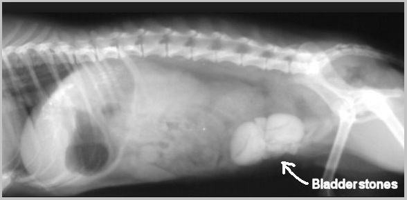
| Canine Hyperuricosuria (HUU) |
| Brief Description: Hyperuricosuria (HUU) means elevated levels of uric acid in the urine. This trait predisposes dogs to form stones in their bladders or sometimes kidneys. These stones often must be removed surgically and can be difficult to treat. |
| How to Test:Many DNA laboratories can test for Canine Hyperuricosuria. |
| Test Type: DNA Cheek Swab |
| Mode of Inheritance: Autosomal Recessive |
| In simple terms: To be affected by Canine Hyperuricosuria (HUU) a dog must have two copies of the affected gene, one from each parent. |
| Clear - Does not carry the affected gene | Is not affected | Cannot pass on the affected gene to offspring |
| Carrier - Carries one copy of the affected gene | Is not affected | Can pass on one copy of the affected gene to offspring |
| Affected - Carries two copies of the affected gene | Is affected | Will pass on one copy of the affected gene to offspring |
| For more information on Canine Hyperuricosuria please click here! |
|
| Progressive Retinal Atrophy (rd1PRA) |
| Brief Description: PRA is a disease of the retina. This tissue, located inside the back of the eye, contains specialized cells called photoreceptors that absorb the light focused on them by the eye’s lens, and converts that light, through a series of chemical reactions into electrical nerve signals. The nerve signals from the retina are passed by the optic nerve to the brain where they are perceived as vision. The retinal photoreceptors are specialized into rods, for vision in dim light (night vision), and cones for vision in bright light (day and color vision). PRA usually affects the rods initially, and then cones in later stages of the disease. |
| How to Test: Many DNA laboratories can test for Progressive Retinal Atrophy. |
| Test Type: DNA Cheek Swab |
| Mode of Inheritance: Autosomal Recessive |
| In simple terms: To be affected by Progressive Retinal Atrophy (rd1PRA) a dog must have two copies of the affected gene, one from each parent. |
| Clear - Does not carry the affected gene | Is not affected | Cannot pass on the affected gene to offspring |
| Carrier - Carries one copy of the affected gene | Is not affected | Can pass on one copy of the affected gene to offspring |
| Affected - Carries two copies of the affected gene | Is affected | Will pass on one copy of the affected gene to offspring |
| For more information on Progressive Retinal Atrophy please click here! |
|
| Hereditary Cataract (HC) |
| Brief Description: Dogs with Hereditary cataracts most commonly present within a few weeks to months after birth with small cataracts that are visible on a veterinary eye exam. Cataracts from this disease will eventually affect the whole lens in both eyes leading to complete blindness between 2-3 years of age. |
| How to Test: Many DNA laboratories can test for Progressive Retinal Atrophy. |
| Test Type: DNA Cheek Swab |
| Mode of Inheritance: Autosomal Recessive |
| In simple terms: To be affected by Progressive Retinal Atrophy (PRA) a dog must have two copies of the affected gene, one from each parent. |
| Clear - Does not carry the affected gene | Is not affected | Cannot pass on the affected gene to offspring |
| Carrier - Carries one copy of the affected gene | Is not affected | Can pass on one copy of the affected gene to offspring |
| Affected - Carries two copies of the affected gene | Is affected | Will pass on one copy of the affected gene to offspring |
| For more information on Hereditary Cataracts please visit here! |
|
| Hip Dysplasia (HD) |
| Brief Description: In dogs, hip dysplasia is an abnormal formation of the hipsocket that, in its more severe form, can eventually cause crippling lameness and painful arthritis of the joints. It is a genetic (polygenic) trait that is affected by environmental factors. |
| How To Test: Most Veterinary Clinics can do the required X-Rays for Hip & Elbow Scoring. The X-Rays are then sent to a Veterinary Radiologist in Australia for Scoring. |
| Test Type: X-rays and scoring by a Veterinary Radiologist |
| Mode of Inheritance: Multifactoral Trait - meaning genetic and environmental factors are involved |
| In simple terms: While it is known that genetics play a huge part in Hip Dysplasia, environmental factors are also the cause or can exacerbate the disease. |
| For more information on Hip Dysplasia please click here! |
|
| Elbow Dysplasia (ED) |
| Brief Description: Canine elbow dysplasia (ED) is a disease of the elbows of dogs caused by growth disturbances in the elbow joint. There are a number of theories as to the exact cause of the disease that include defects in cartilage growth, trauma, genetics, exercise, diet and so on. |
| How To Test: Most Veterinary Clinics can do the required X-Rays for Hip & Elbow Scoring. The X-Rays are then sent to a Veterinary Radiologist in Australia for Scoring. |
| Test Type: X-rays and scoring by a Veterinary Radiologist |
| Mode of Inheritance: Multifactoral Trait - meaning genetic and environmental factors are involved |
| In simple terms: While it is known that genetics play a huge part in Elbow Dysplasia, environmental factors are also the cause or can exacerbate the disease. |
| For more information on Elbow Dysplasia please click here! |
|
| Cardiac Disease |
| Brief Description: Heart disease in dogs, as in people can be either present at birth or acquired, often developing during middle age. Acquired heart disease is more common, affecting many older dogs. Several forms of potentially heritable heart disease have been seen in the American Staffordshire Terrier breed. These include: Subvalvular Aortic Stenosis, Pulmonic Valvular Stenosis, Mitral Valve Dysplasia and others |
| How to Test: There are a number of Veterinary Cardiologists within Australia. |
| Test Type: Auscultation and/or Echocardiography by a Veterinary Cardiologist |
| Mode of Inheritance: Various - Congenital, Genetic |
| For more information on Cardiac Disease please click here! |
|
| Thyroid Disease |
| Brief Description: The thyroid gland controls the metabolic rate of the body. When the gland functions insufficiently, a condition known as hypothyroidism occurs. Dogs that are clinically affected may display one or more of the following clinical signs: weakness, lethargy, weight gain to the point of obesity, skin and coat problems, behavioral abnormalities, and infertility. |
| Test Type: Blood Sample |
| For more information on Thyroid Disease please click here! |
|
| Juvenile Laryngeal Paralysis & Polyneuropathy (JLPP) |
| Note: JLPP is primarily seen in Black Russian Terriers and Rottweilers, however a number of Amstaffs are presenting with this disease. The Staffordshire Terrier Club of America are currently calling for samples from affected dogs to help develop a DNA test for the disease in Amstaffs as the gene mutation is different to those in play in BRT's and Rottweilers. |
| Brief Description: JLPP is a disease that causes deterioration of the nervous system. The longest nerve which controls the muscles of the larynx (voice box) is affected first. This leads to muscle weakness and obstruction of air flow into the lungs after exercise or when the dog is hot. |
| How to Test: Currently there is no test for the Amstaff for JLPP, Researchers are currently working to develop a test. |
| Test Type: In other breeds it is a simple DNA Cheek Swab |
| Mode of Inheritance: Autosomal Recessive |
| In simple terms: To be affected by Juvenile Laryngeal Paralysis & Polyneuropathy (JLPP) a dog must have two copies of the affected gene, one from each parent. |
| Clear - Does not carry the affected gene | Is not affected | Cannot pass on the affected gene to offspring |
| Carrier - Carries one copy of the affected gene | Is not affected | Can pass on one copy of the affected gene to offspring |
| Affected - Carries two copies of the affected gene | Is affected | Will pass on one copy of the affected gene to offspring |
| For more information on Juvenile Laryngeal Paralysis & Polyneuropathy please visit here! |
|


